
The clinical picture of gout
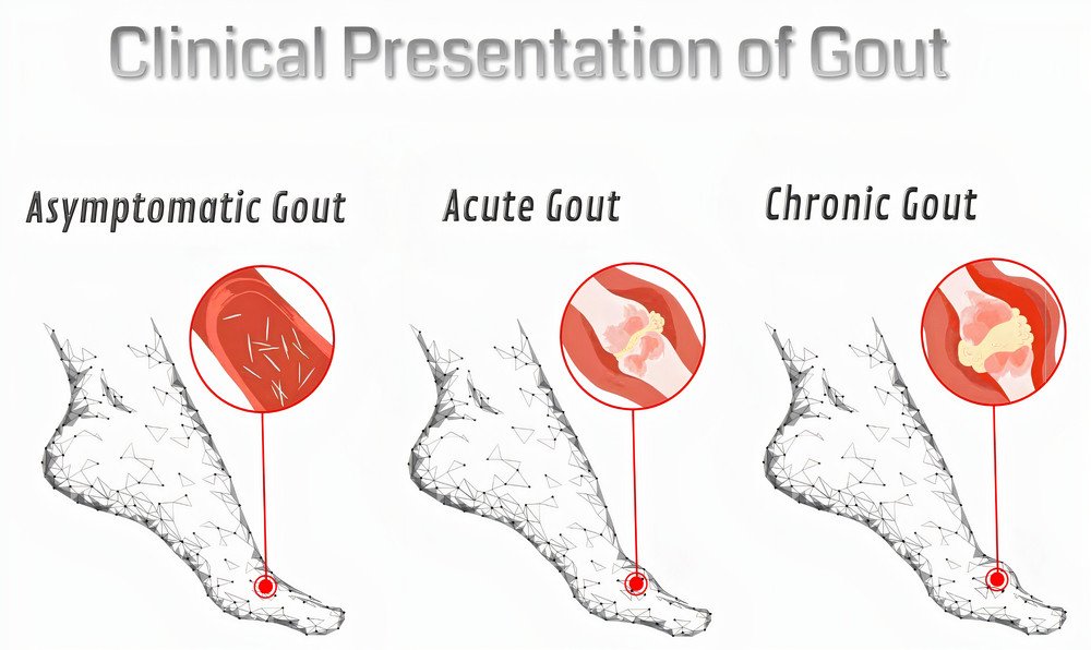
The clinical presentation of gout may be asymptomatic hyperuricemia, acute, or chronic gout. The treatment of gout will depend on the clinical presentation.
1) Asymptomatic hyperuricemia: It means just elevated levels of uric acid without pain or any other symptoms. The majority of hyperuricemic people won’t develop gout.
2) Acute gout: Acute attacks of gout show the following characters in the affected joint:
It is monoarthritis, and it usually affects the metatarsophalangeal joint of the big toe. Sometimes, it may affect the ankle, knee, dorsum of the foot, and rarely the upper limb joints.
The affected joint is painful, hot, red, tender, and with limited movement. The acute attack is so painful, and the pain usually occurs at night and reaches its maximal intensity within hours, which awakens the patient in the early morning. Fever and chills may occur during the attack. Between attacks, the patient is asymptomatic, but during recovery, some itching may occur.
The acute gout is non-erosive and resolves without deformity.
Acute gout may rarely affect multiple joints, such as in leukemia, lymphoma, longstanding gout, chronic renal failure, and post organ transplantation.
3) Chronic gout: In chronic cases, uric acid deposits in the joints (chronic gouty arthritis), kidney (gouty nephropathy), and other tissues (tophi).
- Chronic gouty arthritis shows the following features: It is polyarthritis, asymmetrical, and shows remission and exacerbation. It is erosive and causes joint deformity.
- Gouty nephropathy (urate nephropathy): The presentation of kidney affection depends on the site of uric acid deposition.
Uric acid deposition in renal tubules leads to acute renal failure. In the renal interstitial tissue, it will lead to chronic renal failure. In the urinary tract, it will cause stones, which may lead to urinary tract infections and hematuria. - Tophi: It is hard, painless, yellowish-white, gray nodules that may ulcerate and discharge gray paste material. It occurs in severe hyperuricemia and usually locates on the skin around joints, and extensor surface of the fingers, and ear lobule. It may cause permanent damage to the joint due to bone erosions.
⇒ Now, let’s discuss how your doctor will diagnose you.