
What are the abnormal colposcopic findings?
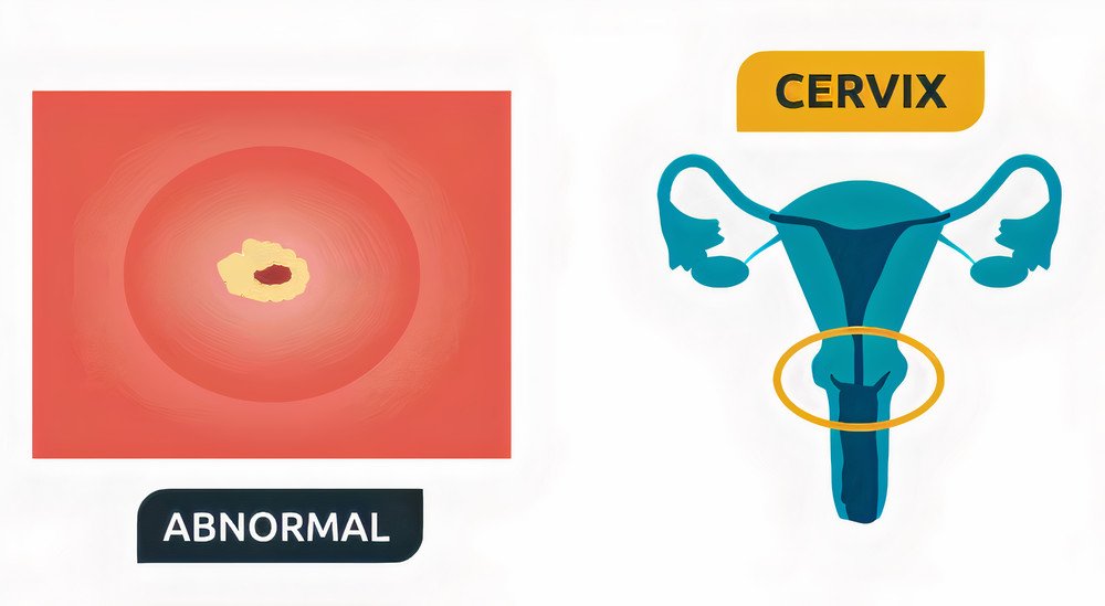
The doctor may see :
- Irregular friable cauliflower Like growths from the cervix with areas of hemorrhage and necrosis ( most probably cancerous).
- Focal white lesions on administering acetic acid with well defined borders and outward extension from the cervix which could be precancerous cells that require further investigation.
- Leukoplakia which are also white lesions raised above the surface that don’t need acetic acid to be seen and require a biopsy.
- Atrophic areas that are wasted away.
- Any abnormal vascular pattern of punctate or mosaic shapes and any abnormally branching vessels like comma shaped or cork screw shaped branches that are suggestive of micro invasion of the sub epithelium.
- Poor iodine staining cells in Schiller’s test that are dysplastic in nature.
Your physician has to describe the lesions shapes and sizes and margins whether it’s taking up all of the four quadrants of the cervix and the direction of its extension into the cervix or away from the cervix and he can also take some images of the lesion to reassess them later.
The doctor may also get unsatisfactory results like the inability to visualize the transformation zone (TZ) to its upper limit as it could be shifted upward to the endocervix normally as in menopausal women by the effect of different hormones.
Then your doctor will ask for either endocervical curettage or a biopsy to be taken in the same setting if the lesion requires further investigations but if the lesion is diagnosed easily he may try to treat it by excision or may decide to take time for following its progression.