

Breast cancer is one of the leading causes of female mortality around the world. It is one of the most common cancers diagnosed in women, only second to invasive cancer of the skin. The well known statistic that one in eight women will get invasive breast cancer is a halting reminder of just how common this disease is. Another figure that drives this point home is that one third of cancers that are diagnosed in women are breast cancers.
When a woman feels a lump or some pain in her breast, it is obvious that something is wrong. But the benefit of radiological studies is that they can detect a disease before it causes any symptoms. The most commonly used and overall most useful imaging study for the detection of breast cancer is the mammogram. It can detect most breast cancers at least one year before they can be felt by the patient or her physician. It is relatively cheap, widely available, and very accurate. Along with other advances in treatment modalities and increased public awareness, breast mammography has contributed significantly to the reduction of mortality rates of breast cancer. Since instituting breast cancer screening using this test, the mortality rate has sharply diminished. Between 1990 and 2010: the mortality rate from this disease has dropped by an estimated 34%.
Mammography is a very commonly used imaging study. It is reportedly performed about 48 million times every year in the United States. This article will cover what exactly a mammogram is, the use of mammography for screening purposes, 3D mammography or digital breast tomosynthesis, the amount of time needed to perform a mammogram, whether or not mammograms cause any pain or discomfort, the most appropriate time to get a mammogram, diagnostic mammography, the use of mammography in detecting breast cancer, what constitutes an abnormal mammogram, the personal preparations you need to do before a mammogram is performed, mammography recommendations, mammogram results, and how much money a mammogram costs in the US with and without insurance.
What is a mammogram?


Etymology: “Mammo” meaning breast, and “-gram” meaning a recording.
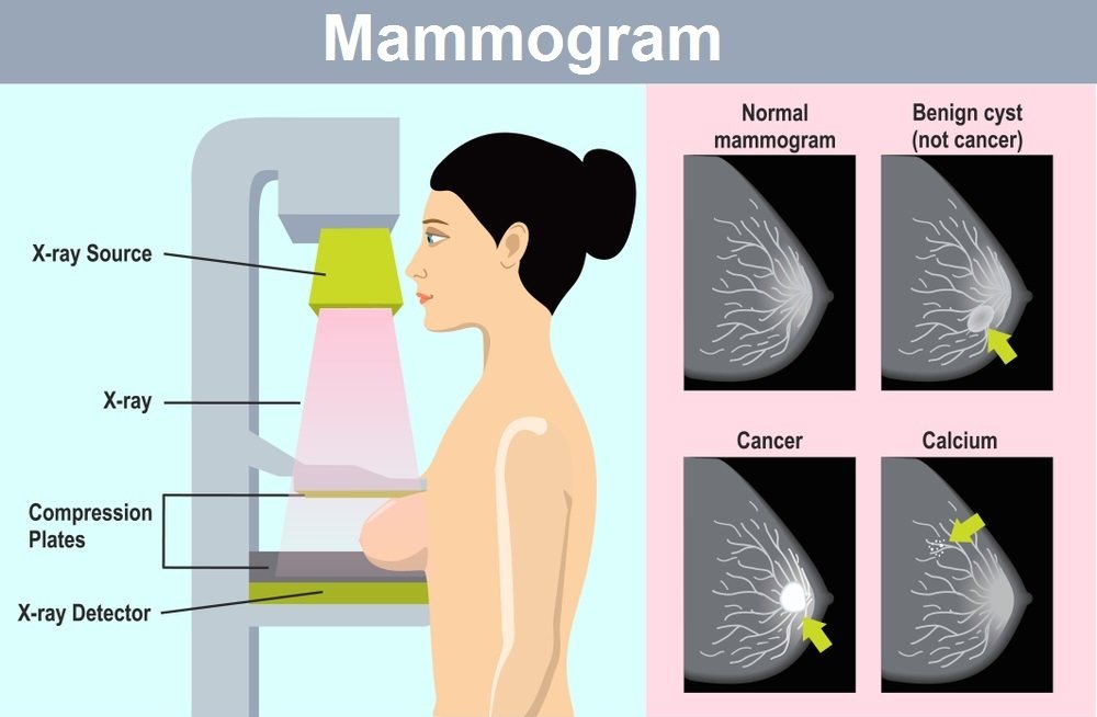
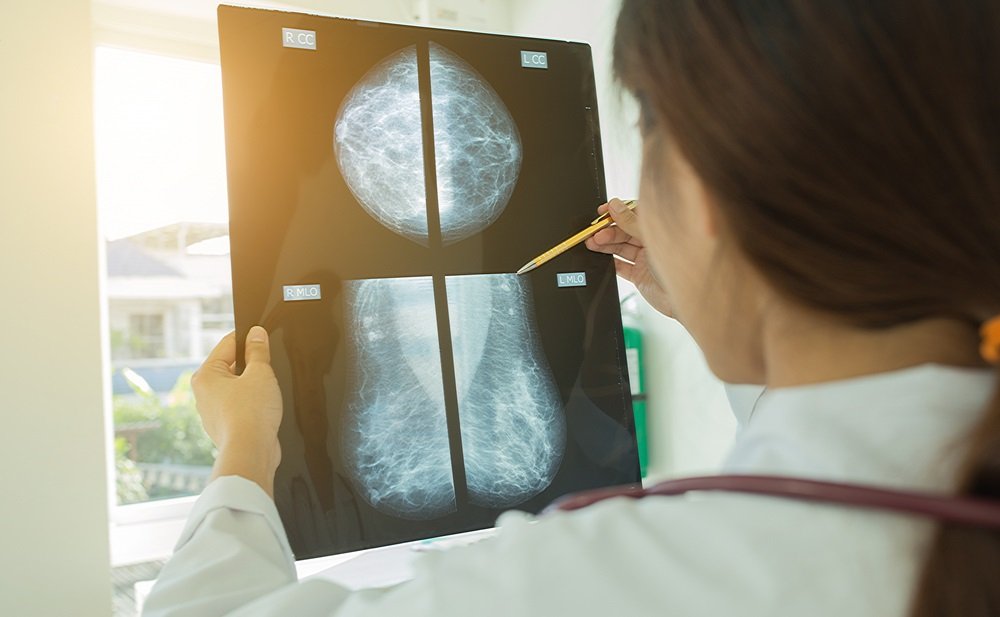
A mammogram is a special imaging study that uses X-rays to check the breast for any pathology. A machine produces the X-rays, which penetrate the breast tissue. During this process, the more dense the tissue, the more rays are absorbed. This is why in conventional radiographs: bones, which are very dense, appear white, while the lungs, which are far less dense, appear black. This principle is the basis of a mammogram’s ability to detect abnormal structures in the breast. Typically the xrays are shot from two different angles at least: the craniocaudal and the mediolateral oblique. This is useful because if a radiologist sees two or more overlapping structures, he/she will need to look from another angle to assess them properly.
For context: Xrays are a form of electromagnetic radiation. They were discovered in the 19th century by the German physicist Wilhelm Röntgen. He named them xrays to signify their elusive and unknown nature. The famous story which paved the way for the medical usage of xrays is that Röntgen photographed his wife’s hand with an xray machine. It was the first xray image taken of a human, and it showed the bones in unprecedented detail, in addition to the wife’s ring.
Technically speaking, a mammogram can be taken in two formats: digital mammography and film mammography. Film mammography pastes the xray image onto a physical film, and requires a proper light source to read well. Digital mammography creates an image on a computer. This type of radiography offers the advantages of using a lower dose of radiation, having higher contrast resolution, and giving the radiologist the ability to zoom into specific parts and change the contrast and brightness as well. Since digital mammography is more accurate with dense breasts, it is the better modality to use in younger women who have not yet hit the menopause period.
Continue reading to learn about mammogram screening now.
Mammogram screening


In medicine, screening refers to the testing of a person who doesn’t show any signs or symptoms in order to detect a disease in its early stages before it starts manifesting. Thus screening mammography is the type of mammography used in women who do not suffer from any signs or symptoms of breast disease, such as breast pain, a lump or swelling in the breast, nipple discharge, or any other physical deformity. This allows early detection and thus early treatment, which is associated with a better prognosis.
Mammography has contributed to a 36% decrease in breast cancer mortality rate from 1989 to 2012, and a lowered risk of death is not the only advantage of the early detection of a breast lesion. The same breast lump that would have needed a minimally invasive surgery to remove if it had been discovered early may need a much more disfiguring operation if discovered late when it has increased in size and encroached upon other thoracic structures. For example, a breast mass may be too small to feel during a clinical examination but may show up on a screening mammogram. When women above 40 years of age get annual mammograms, the type they’re getting is the screening mammogram.
It should be mentioned that women who had underwent breast reconstruction surgery and have lost all underlying breast tissue need no screening mammograms. Clinical examination of these reconstructed breast is the preferred method of screening. Regarding pregnant women: The risk of radiation exposure to the foetus actually outweighs the benefits of getting screening mammograms regularly, which is why pregnant women only get diagnostic mammograms.
3D mammogram


Also known as digital breast tomosynthesis, this is a special type of screening mammography approved by the Food and Drug Administration FDA in 2011, which creates a 3-dimensional image of the breast tissue by using multiple xray images taken from multiple angles by a rotation xray beam. The advantages of this type of imaging include the detection of slightly more breast cancers than two dimensional mammography. It also limits the need for a follow up mammogram taken from a different angle as it visualises the breast from all angles.
However, since 3 dimensional mammography takes multiple xray images, it may expose the patient to more radiation than a conventional mammogram. Digital breast tomosynthesis is also slightly more expensive than two dimensional mammography. The bottom line is that there isn’t enough evidence to prove that 3D mammography is necessarily better than 2D mammography when it comes to the mortality rate, but that the new technology shows promise. One study, which compared digital breast tomosynthesis with two dimensional mammography, showed that the former was much more accurate than the latter.
How long does a mammogram take?


The procedure takes about 10 to 20 minutes for a screening mammogram. A diagnostic mammogram may take a few minutes more because it uses more angles.
Do mammograms hurt?
During a mammogram, the breast must be compressed between two curved acrylic plates. This is done to reduce the thickness of the breast tissue that the xrays will pass through as much as possible, and to prevent any unnecessary movement and thus any motion blur, allowing for a clearer image. This is necessary even in male subjects, who use smaller plates.
Some women do experience some discomfort or pain because of this, but most tolerated it well. The level of discomfort varies from person to person, and depends on factors such as the timing of the procedure in relation to the menstrual cycle. Taking this particular factor into account, the most appropriate time to schedule a mammogram is the week following the end of the period. Other factors affecting the level of discomfort include the size of the breast, the experience level of the technician, and the level of anxiety of the patient. A minority of patients may experience some soreness after the procedure but most don’t. If you are worried about this aspect, taking an over the counter painkiller before getting the mammogram may help.
When to get a mammogram


Different medical organisations have different opinions concerning when a woman should start getting screening mammograms. What is definitely agreed upon is that women between the ages of 50 and 69 need to get regular mammograms. The controversy concerns women who are younger or older than this age bracket.
For a woman of average risk, mammogram recommendations are as follows: The National Comprehensive Cancer Network, The American College of Surgeons and the American College of Radiology advise women to start getting annual mammograms beginning at 40 years of age. On the other hand, The American Cancer society advises starting at 45 years, with the option to start between 40 and 44. The ACS recommends getting annual mammograms from 45 to 54 years of age, then after reaching 55 years: the woman should switch to getting one radiograph every two years, or stick with annual testing if she so desires. Emphasizing mammography’s superior capacity to detect breast cancer, the ACS makes no recommendation for the use of clinical examination to screen for breast cancer, and advises women to keep getting mammograms as long as they have good health and a life expectancy of ten or more years.
Meanwhile, The US Preventive Services Task Force, after combing through the results of eight randomised controlled trials, advocates for starting at 50 years and getting a mammogram every two years until 74 years of age, again with the option to start at 40 years if the patient thinks it best to do so. Most women in general begin at 40. Some authorities recommend that women with a first degree relative who had been diagnosed with breast cancer should start getting mammograms 10 years prior to the age at which the diagnosis of the relative was made. Talk to your physician to determine the most appropriate time to start getting mammograms, which depends on your particular family history and risk factors.
Diagnostic mammogram


As opposed to a screening mammogram, a diagnostic mammogram is done for patients who have signs or symptoms of breast disease. These include breast pain, nipple discharge, a lump or swelling, or any other visible deformity of the breast. If a screening mammogram shows abnormal findings, a diagnostic mammogram is usually ordered to evaluate them further.
As mentioned above, diagnostic mammograms entail taking more images then normal screening mammograms, allowing for a more detailed assessment of the breast tissue. The conventional angles used in any mammogram are the craniaocaudal and the mediolateral oblique views. The additional angles include “Cleavage view”, “rolled view” and “tangential view”. This is why diagnostic mammography is more expensive and entails slightly more radiation exposure than screening mammograms.
Mammogram prep


The steps you should take before getting a mammogram are as follows:
- Educate yourself. The more you know about the procedure, the less anxious you will feel. Making sure you know the advantages, risks, pricing, duration, and all other relevant facts will make the procedure go much more smoothly.
- Pick a suitable date. As mentioned above, the most appropriate time to select in order to minimize the discomfort felt during a mammogram is one week after your period ends. If pain is a concern, taking an over the counter pain medication beforehand may help reduce the discomfort and soreness. If you experience excessive discomfort during the procedure make sure to tell the technician.
- Avoid using the following products before going to the medical facility: Some personal care products may show up on the xray and jeopardise the mammogram. These include deodorants, creams, lotions, and perfumes.
- You will be asked to uncover your upper body, so it is advisable to wear a two piece outfit to the medical facility.
- If this is not your first time getting a mammogram, make sure to bring your previous ones so that the radiologists can compare them.
Mammogram results


As detailed above, a patient who doesn’t show any signs or symptoms of breast disease should undergo a screening mammogram starting at a certain age. If no positive findings are discovered, the patient is sent home and no additional procedures are required. This should not discourage patients from undergoing annual mammograms, as breast disease can arise at any time. If, however, an abnormal finding is discovered, a diagnostic mammogram is usually ordered to analyze the pathology in more detail.
Abnormal mammogram

Mammograms are made when low energy X Rays are passed through the breast tissue. Depending on the density of the different areas of the breast, the xrays are absorbed in different patterns. The denser the tissue, the more xrays it absorbs. A dense portion of tissue appears more white on the xray film, while a darker part indicates that it has less density.
In general, the breast becomes less dense as the patient’s age increases. Breast implants may make it more difficult to read a mammogram as they appear opaque on the film. In addition, breast implants make it difficult to compress the examined breast during the procedure, further decreasing the quality of the taken radiograph. This is why the best imaging study for women with breast augmentation is the MRI, not the mammogram.
There are a number of abnormal findings that can be detected on a mammogram other than a tumour, and they are usually much less dangerous and more common. These include calcium deposits or calcifications, which are collections of calcium particles in a certain area usually due to inflammation or trauma. Calcium deposits often increase with age. another finding is that of a cyst which is a thin walled sack filled with liquid. Yet another finding is a fibroadenoma, which is a benign tumour that usually appears in young adults. They are usually small, round and easy to move . Coming back to the topic of density, high breast density is associated with a slightly increased risk of breast cancer.
Breast cancer mammogram

Since a breast lump or mass usually has a higher density than the surrounding tissue, the radiologist primarily looks for high density white spots. If one or more such findings are discovered, additional imaging studies may be ordered such as an ultrasound or magnetic resonance imaging MRI.
One drawback of imaging studies in general is that they cannot determine whether a breast mass is benign or malignant. For this, a tissue biopsy is often needed. The key distinction between benign and malignant tumours is that malignant tumours can spread to the rest of the body, a process known as metastasis, while benign tumours do not metastasize. This does not mean that benign tumours are harmless, they can cause discomfort and cosmetic disfigurement, but they have a much lower mortality rate. To decide if a tumour is benign or malignant, it is necessary to examine a portion of it under a powerful microscope. The process of extracting a part of the tumour for diagnostic purposes is called a tissue biopsy. It is often done using a needle after applying an anaesthetic. Thus, a full diagnosis of a breast mass is reached by using what medical professionals call a triple assessment. This begins with a clinical examination of the patient’s breast, then one or more radiological studies, and finally a tissue biopsy.
It is not always easy to differentiate a benign finding from a more dangerous lesion, which hich is why we recommend bringing any previously taken mammograms when you go to get a new one, so that the radiologists can compare the old and the new.
Risks of mammography

All medical procedures carry some risks and mammography is no exception. The benefits of getting a mammogram usually outweigh the risks, but a brief discussion of the less desirable aspects of a mammography is needed to paint a complete picture.
Since a mammogram depends on using the electromagnetic radiation we call xrays, it is inevitable that some radiation exposure will occur. This is usually of a very low dose which shouldn’t cause any concern. To put this in perspective, the radiation that a woman is exposed to yearly from background environmental sources is more than that received during a mammogram. The main situation in which radiation exposure needs to be taken into account Is when examining a pregnant woman. The foetus is particularly sensitive to radiation, especially in early pregnancy. This is why women who are pregnant do not get regular screening mammograms but only diagnostic mammograms. Women with the well known genetic mutations BRCA1 or BRCA2 are also more at risk for genetic damage caused by the radiation.
As mentioned above, some factors may make the interpretation of a mammogram difficult, such as increased breast density or breast implants. This raises the possibility that the patient may be diagnosed with a lesion that isn’t actually there, which is called a false positive, or be told that there are no abnormal findings while in fact some lesions are present, which we call a false negative. Since younger women have more dense breasts, , they are more likely to receive an inaccurate diagnosis. This fact contributes to the controversy around the validity of recommending annual mammograms for women less than 50 years of age.
Along the same vein, some types of breast cancer may not show up on a mammogram. If some breast disease is causing symptoms but does not appear on the mammogram, additional radiological studies may be ordered.
How much does a mammogram cost in the USA?


Before discussing pricing, it should be emphasized that mammograms are without a doubt worth the financial cost. This is a relatively cheap investigation with the potential to save a life. If we remember that one in eight women are afflicted with and may potentially lose their lives because of breast cancer, getting annual mammograms becomes a no brainer.
The financial aspect of mammography depends on whether or not the patient has insurance. It should also be added that 3 dimensional mammography costs a bit more than a standard 2 dimensional mammogram, and insurance may not necessarily cover the former.
For those with insurance: Medicare, Medicaid, and most insurance companies cover screening mammograms for women 40 years or older with no out of pocket costs.
For those without an insurance: The price varies according to the selected facility, so it helps to shop around. The prices may vary from $80 up to $200.