

Peripheral vascular disease is a vascular condition that impairs blood flow through the peripheral circulation, which includes the blood vessels that carry blood away from the heart to the peripheral tissues rather than the heart and brain, such as the upper and lower extremities.
In peripheral vascular disease, peripheral blood vessels narrow due to a variety of causes, which decreases blood supply to the peripheral tissues. Low blood supply means low oxygen supply and ischemia. Without enough blood and oxygen, body cells can’t perform their function well, and they may die if this ischemia prolonged, which leads to a variety of symptoms depending on the affected part. The peripheral vascular disease usually affects the legs, and its leg symptoms range from pain to gangrene, which may require amputation if left untreated. Thus, early diagnosis and proper treatment are essential to avoid tissue loss and disability.
Peripheral vascular disease affects both sexes and more common among older people. According to the center for disease prevention and control (CDC), it affects 12%-20% of people older than 60 years old. It is a potential cause of death among people older than 60 years and diabetics, and most people with PVD are asymptomatic. The good news is that we have effective management plans that can prevent a lot of complications and improve the quality of life for these patients.
The peripheral vascular disease usually affects the arteries; thus, peripheral vascular disease (PVD) is another name for peripheral artery disease (PAD).
we will know more about the peripheral vascular disease by answering the following questions:
- What are the causes and risk factors of peripheral vascular disease?
- What is the pathophysiology of this disease?
- What are its symptoms?
- How do physicians diagnose it?
- How can physicians treat it?
Causes and risk factors of peripheral vascular disease


The main cause of peripheral vascular disease is atherosclerosis, which means deposition of fatty material (atherosclerotic plaques) in the wall of blood vessels. With time, these plaques reduce the blood flow through the affected vessels and may block it.
Risk factors of atherosclerosis raise the risk of peripheral vascular disease, and they include non-modifiable and modifiable risk factors, as follows:
A) Non-modifiable risk factors:
- Age more than 50 years old
- Males have a risk two times more than females.
- Positive family history
- Type A personality
B) Modifiable risk factors:
- Smoking is the most significant risk factor, either first or second-hand smoking. Every cigarette raises the risk of peripheral vascular disease more and more. 90% of patients with peripheral vascular disease are smokers.
- Diabetes mellitus increases the risk of all cardiovascular diseases, including peripheral vascular disease. It also increases the risk of complications, such as gangrene.
- Hypertension is a common risk factor for atherosclerosis because it damages the wall of the blood vessels.
- Dyslipidemia means high levels of LDL cholesterol (bad cholesterol), low levels of HDL cholesterol (good cholesterol), and high triglycerides in the blood, which raises the risk of atherosclerosis.
- Obesity and overweight
- A sedentary lifestyle with minimal or no physical activity raises the risk of atherosclerosis and all cardiovascular diseases.
Other causes of peripheral vascular disease include:
1. Vasculitis: Inflammation of blood vessels narrow their lumen and impairs blood flow.
2. Injury of blood vessels as in accidents
3. Some inherited structural disorders cause the narrowing of blood vessels.
4. Infections such as syphilis
5. Cold temperature can cause vasospasm and decrease the blood flow to the peripheral tissues.
6. Stress: Chronic stress increases the sympathetic tone, which leads to vasoconstriction, raises blood pressure, and reduce blood flow through the vessels.
⇒ Now, we will discuss how peripheral vascular disease occurs.
Pathophysiology of peripheral vascular disease


In peripheral disease, peripheral tissues don’t get enough oxygen and nutrients to survive and do their functions. Ischemic cells respond to the lack of oxygen by releasing adenosine, which is a signaling molecule that affects nerves and causes pain. Severe lack of oxygen can cause tissue death (gangrene).
According to how the peripheral vascular disease occurs, we can classify it into two types:
1. Organic PVD: It means a reduction of blood flow due to abnormality in the blood vessel structure, such as in atherosclerosis, where the plaques interfere with blood flow.
2. Functional PVD: It means narrowing of the blood vessels due to constriction of the smooth muscle in its wall. Under normal conditions, the nervous system affects the blood vessels, either constrict or dilate it, but peripheral vascular disease aggravates thee vasoconstriction, such as in stress and cold temperature.
Clinical presentation of the peripheral vascular disease depends on:
- The affected blood vessel
- Sudden ischemia (embolus) or thrombus developed gradually over time
- Presence of collateral circulation in the site of occlusion
⇒ Now, let’s discuss the clinical picture of peripheral vascular disease.
The clinical picture of peripheral vascular disease


Most people with peripheral vascular disease are asymptomatic. Symptomatic patients almost always have lower limb problems. The most common symptom is a severe dull cramping leg pain during walking and exercise, which is called intermittent claudication. This pain occurs during activity because the muscles require more oxygen while rest reduces the oxygen demand by muscles and relieves the pain. The pain site depends on the affected vessel; it may be in the buttocks, thighs, calves, or feet, but it is more common calves. It also may occur in one or both legs. Other symptoms may include leg fatigue, heaviness, and tightness.
Many people ignore these symptoms as an expected sign of aging, which may lead to worsening of the disease and dangerous complications. As the disease progresses, there is a further lack of oxygen supply to the tissues that may reach the degree that doesn’t meet the tissue needs at rest, which causes critical limb ischemia. critical limb ischemia leads to the following:
- The pain worsens to be at rest also and becomes severe enough to restrict any movement.
- Cold legs and color changes to be pale or blue
- Wasting of the leg muscles
- Numbness and tingling in legs
- Loss of hair in legs
- Thick and opaque nails
- Leg ulcers that don’t heal over the pressure sites, such as ankles
- Erectile dysfunction and impotence
- Severe ischemia impairs the healing of wounds, and unhealed wounds may lead to fatal infections of the bone or bloodstream.
- Finally, we can end in gangrene (tissue death), which may require amputation of the dead part to avoid the loss of the whole limb and septicemia.
Peripheral vascular disease can be a part of cardiovascular disease that may affect the heart and brain and cause fatal events like stroke and myocardial infarction; stroke incidence is higher in patients with peripheral vascular disease. Thus, if you felt any symptom of peripheral vascular disease, you should see your doctor and don’t ignore it. Early diagnosis and management of peripheral vascular disease prevent further damage to blood vessels and more tissue damage, which reduces the risk of tissue loss and life-threatening complications.
⇒ Now, let’s discuss how your doctor will diagnose you.
Diagnosis of peripheral vascular disease


First, when you tell your doctor about your symptoms, he will ask you about your medical history, such as smoking, your lifestyle, and if you have diabetes, hypertension, or any disease.
After history taking, the doctor will examine you to consider a specific diagnosis. The physical examination will include things, such as:
1) Ankle/brachial pressure index

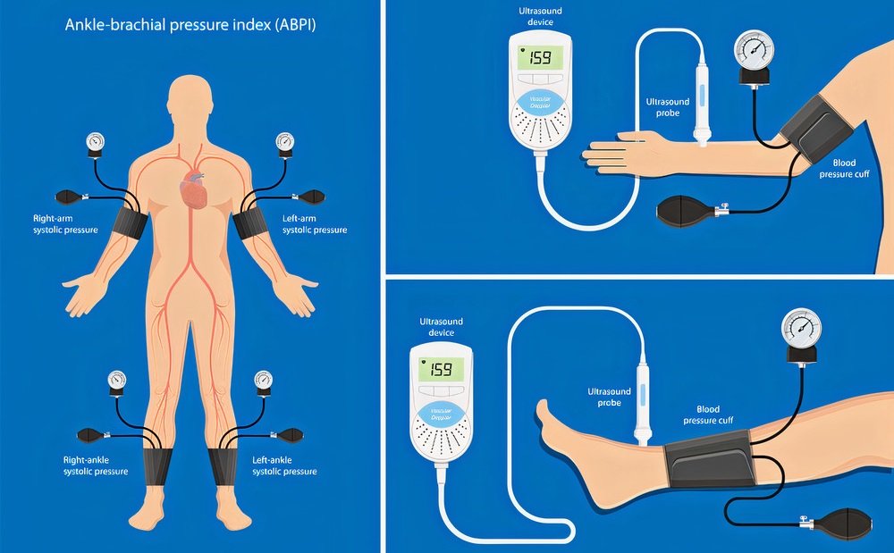
It is the first test that after suspicion of peripheral vascular disease. The doctor will measure your blood pressure in the arm and leg. Then he will take the ratio between the leg and arm pressures. If this ratio is 0.9:1.2, it is within the normal range. If the result is less than 0.9, it will suggest peripheral vascular disease. According to the ankle/brachial index, we can classify the peripheral vascular disease into mild (0.7:0.9), moderate (0.5:0.7), and sever (less than 0.5). If the result is higher than 1.4, it indicates noncompressible arteries.
Noncompressible arteries mean hardening of their wall due to calcification, which occurs in diabetic patients. These results (>1.4) are false-negative results, which require further investigations because these patients often have severe peripheral vascular disease and a higher risk of death from cardiovascular disease.
2) Buerger’s test

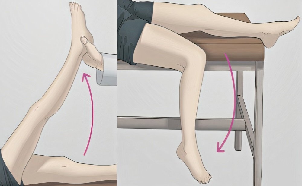
The idea of this test is to see if there is pallor after leg elevation and assess if there is arterial insufficiency. In normal people, the legs remain pink after being raised by 90 degrees (vascular angle). In peripheral vascular disease, the affected leg becomes pale after elevation by 30 degrees for 30 or 60 seconds, and in severe ischemia, it occurs after 15 degrees or less.
Also, during the physical examination, the doctor will check things, such as:
- Muscle wasting
- Compare the affected limb temperature with the other healthy limb
- Hair loss
- Thick and opaque nails
- Examine the sensation in the affected leg to assess if there is tissue loss
After history taking and physical examination, your doctor may require further investigation to confirm his suspicion about peripheral vascular disease. These investigations include:
3) Treadmill exercise test


Your doctor will request this test if he still suspects peripheral vascular disease despite a normal ankle/brachial pressure index. In this test, the doctor measures your blood pressure in the arms and legs before the exercise then he will ask you to exercise (on the treadmill) till claudication occur. Then he will retake your blood pressure in the arms and legs and recalculate the ankle/brachial pressure index.
4) Imaging
Many imaging techniques can help in the diagnosis of peripheral vascular disease through the visualization of blood vessels and blood flow through it. These techniques include:
A) Doppler ultrasonography: It is a non-invasive technique that provides an excellent way to assess the blood flow through the vessels. In this technique, the computer uses high-frequency ultrasound waves to take a picture of blood vessels.
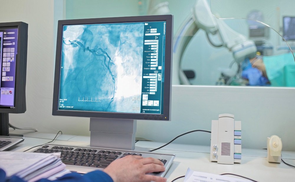
B) Angiography (x-ray of blood vessels): In this technique, the radiologist injects a contrast dye into a leg artery to highlight the blood vessels on the x-ray, which helps to detect blockage or narrowing. This technique has disadvantages, such as being invasive, using dye makes it contraindicated in renal failure, and taking much time to do it makes it hard in acute cases when there is a need for immediate treatment.
C) Magnetic resonance angiography (MRA): Magnetic resonance imaging technique uses the power of the magnetic field to provide highly detailed for the internal structures like blood vessels. Magnetic resonance angiography saves the patients from adverse effects of exposure to irradiation besides its high accuracy. Disadvantages include the high cost, which limits its availability.
D) Computed tomography angiography (CTA): This technique depends on the x-ray to image specific areas of the body. We can do it, either by contrast dye or not. Contrast dyes provide better imaging, but we can’t use it in renal failure. Despite the disadvantage of exposure to irradiation, computed tomography has advantages that make it so useful in the emergency, such as no availability and time obstacles as MRA and its high accuracy.
Risk factors are an essential element in the management plan of peripheral vascular disease; thus, your doctor may require investigations to evaluate them, such as:
- Blood glucose for diabetes if he suspects it. If the patient has diabetes, the doctor will order “Hb-A1c” to evaluate the management plan of diabetes.
- Body mass index (BMI) for overweight or obesity
- Lipid profile for dyslipidemia: low-density lipoprotein (LDL), high-density lipoprotein (HDL), and triglycerides (TGs)
- Echocardiography and electrocardiography (ECG) to assess the cardiac functions
Edinburgh Claudication Questionnaire


This questionnaire can be useful for both physicians and patients. It consists of six questions, as follows:
- Do you feel pain or discomfort in your leg(s) on walking?
- Positive response → Yes
- If the response is No, don’t complete.
- Does the pain ever begin when you are standing or sitting?
- Positive response → No.
- Do you get this pain if you walk up or hurry?
- Positive response → Yes
- Do you get this pain when you walk at a usual speed on a level?
- The response depends on the severity of the condition.
- What happens if you stand still or sit?
- Positive response → pain disappear in 10 minutes or less
- Where do you feel the pain?
- Positive response → Typical claudication => in calves
Atypical claudication → in buttocks or thighs.
To say there is peripheral vascular disease, you should have positive responses to all questions as above. The accuracy of this questionnaire reaches 90% in people with peripheral vascular disease.
After diagnosis, If the doctor confirmed the peripheral vascular disease, assessed the severity of the condition, and evaluated the potential risk factor, he would put a plan to manage this condition and prevent further worsening.
⇒ Now, let’s discuss the treatment of peripheral vascular disease.
Treatment


The goals of the management plan are:
- Control the symptoms
- Improve the quality of life
- Prevent life-threatening complications
- Avoid tissue loss (amputation) as possible
The management plan depends on the severity of the condition, and it includes lifestyle modifications, medications, interventional procedures, and surgery.
1) Lifestyle modifications


Your doctor will recommend you modify some aspects of your lifestyle to control the risk factors, which reduce the symptoms and prevent worsening of the disease. These modifications include:
1. If you were a smoker, you should stop smoking. Smoking cessation reduces disease progression, reduce the symptoms, reduce the risk of all cardiovascular and cerebrovascular diseases, and improve your overall health status. It isn’t late to stop even after diagnosis.
2. Exercise: Regular exercise reduces the risk of all cardiovascular diseases and improves the quality of life in both healthy people and patients. In peripheral vascular disease, regular walking opens the collateral vessels (small alternative vessels) in the affected area, which improves the blood supply and reduces the frequency of the intermittent claudication.
3. Keep your weight within the healthy range. If you were overweight or obese, you should ask a nutritionist to give you a suitable program to reduce your weight to a healthy range.
4. Keep your food healthy and avoid fast food and saturated fats.
5. Avoid stress by using any stress-reduction technique suitable for you.
If you were hypertensive, you should follow your doctor’s instructions to keep your blood pressure within the safe range. It is the same in diabetes; you should follow your doctor’s instructions and keep your blood glucose within a safe range. Also, diabetic patients should regularly examine their feet.
2) Medications


Doctors use drugs in peripheral vascular to control the underlying causes and improve perfusion.
Doctors prescribe medications for underlying causes, such as hypertension, diabetes, or vasculitis. Also, in dyslipidemia, doctors will prescribe lipid-lowering agents -such as statins- that reduce LDL cholesterol levels, which reduces the disease progression.
To control the symptoms and improve perfusion, doctors can prescribe some drugs, such as:
- Antiplatelet drugs, such as aspirin and clopidogrel: These drugs prevent platelet aggregation and clot formation.
- Cilostazol (Pletal): This drug prevents platelet aggregation and causes vasodilation, which improves blood flow.
- Anticoagulants, such as heparin and warfarin: These drugs inhibit coagulation factors, which prevents clot formation and improves blood flow.
These drugs prevent clot formation but don’t remove existing clots. Thus, in cases of acute ischemia, doctors can use thrombolytic agents to remove existing embolus. These drugs should be used only in the hospital, by the doctor, and within 4:8 hours from the symptoms.
3) Interventional procedures


Angioplasty (percutaneous transluminal angioplasty): It is a non-surgical procedure, in which, the physician inserts a catheter with a balloon attached to it through a large artery, such as the femoral artery, to reach the narrowed artery then inflate the balloon and dilate the artery. The doctor could set a stent inside the artery through the catheter to maintain patency if he wanted. Also, he can use the laser through the catheter to dissolve a clot. In acute ischemia, he can apply the thrombolytic therapy through the catheter.
Atherectomy is a subtype of angioplasty, in which the physician uses a cut blade through the catheter to remove the plaque.
- The doctor judges the need for angioplasty, according to:
- The site of the narrow or blocked artery
- The number of obstructed vessels
- The severity of the condition
- Angioplasty has advantages over surgical procedures, such as:
- Being less invasive (No major surgical incisions)
- Avoidance of general anesthesia and its side effects and risks; local anesthesia is enough
- Being a one-day procedure allows you can leave the hospital on the same day and return to your normal activities rapidly.
Angioplasty may have some side effects (such as injury of the catheter insertion site and damage of the artery of insertion), but these complications are so rare.
4) Surgery


Your doctor may decide a need for bypass surgery in many situations, such as if most of the vessel is narrow or blocked, and if there are multiple areas of narrowing. Bypass surgery requires general anesthesia and a long hospital stay. In bypass surgery, the surgeon uses a graft (either part from a body vessel -like the greater saphenous vein- or synthetic vessel) to bypass the narrowing or blockage. The surgeon sutures the two ends of the graft before and after the obstruction, which allow the blood to reach the ischemic part and supplies the required oxygen and nutrients.
In peripheral vascular disease, the doctor will refer you to a surgeon for bypass surgery, only if your condition is severe and there is a high risk for tissue loss, and when the lifestyle modification, medication, and angioplasty failed to control the condition.
Amputation: In the case of gangrene, your doctor will refer you to a surgeon to remove the dead tissue to avoid further tissue loss and septicemia (spread of bacterial toxins from the dead tissue to the blood), which is a fatal condition.
Finally, peripheral vascular disease is a preventable disease; you can protect yourself by some lifestyle modifications, as we discussed before. It is never late to begin this modification; getting a healthy lifestyle can control the symptoms, improve the quality of life, and even reverse the condition. Don’t consider the symptoms of peripheral vascular disease a sign of aging and ignore it, especially if you have risk factors, but see your doctor and take his advice. Remember that the earlier the diagnosis, the more successful the management plan.